How To Treat Bursitis Of The Foot
Overview
Heel Bursitis is a condition where one of the bursae at the back of the heel becomes swollen, inflamed and painful. A bursa is a fluid filled sac that cushions muscles, tendons and joints. There are 3 main types of bursitis associated with heel bursitis. These include Retro-calcaneal bursitis, Achilles bursitis, and Sub-calcaneal bursitis. The locations of the 3 bursae are: the insertion point of the Achilles tendon at the back of the heel (retro-calcaneal bursa), between the Achilles tendon and the skin at the back of the heel (Achilles bursa), and the bottom of the heel (sub-calcaneal bursa).
Causes
Bursitis occurs when the bursae become irritated or infected, often causing pain on movement. When infection is involved, medical intervention is necessary to fight the underlying infection and prevent it from spreading, when infection is not involved, prompt medical attention can prevent the condition from becoming worse over time.
Symptoms
You might have Retrocalcaneal Bursitis if you notice any of the following symptoms. You have pain or tenderness at the back of the heel where the Achille's tendon attaches. Have swelling near the attachment of the tendon to the heel bone. You have noticed a slowly growing bump on the back of the heel. The back of the heel turns red after getting rubbed in shoes. The back of the heel hurts worse when you run, walk up hill or wear high heels.
Diagnosis
Your doctor will take a history to find out if you have the symptoms of retrocalcaneal bursitis. By examining your ankle, he or she can generally tell the location of the pain. The physician will look for tenderness and redness in the back of the heel. The pain may be worse when the doctor bends the ankle upward (dorsiflex), as this may tighten the achilles tendon over the inflamed bursa. Alternatively, the pain may be worse with toe rise, as this puts stress on the attachment of the achilles tendon to the heel bone. Imaging studies such as X-ray and MRI are not usually necessary at first. If initial treatment fails to improve the symptoms, these studies may be obtained. MRI may show inflammation.
Non Surgical Treatment
One of the most effective treatments for infracalcaneal bursitis is to temporarily avoid weight-bearing activities that put stress or strain on your heel bone. PRICE (protection, rest, hot/cold contrast compresses, compression, and elevation) is another good acute management technique for this foot problem. Changing your footwear is an effective long-term prevention and treatment tool for infracalcaneal bursitis. Footwear that possess a flat support base, a sufficiently wide toe box to accommodate natural toe splay, and a flexible sole are best for preventing and managing infracalcaneal bursitis. An integrated approach to this problem usually involves the use of padded socks that help reduce pressure, friction, and inflammation in your affected area. Natural anti-inflammatory agents can also be helpful in quelling inflammation, reducing pain, and improving treatment times for infracalcaneal bursitis. In rare cases, more aggressive treatment methods may be required, such as cortisone injections or surgery to drain the bursal sac.
Surgical Treatment
Surgery. Though rare, particularly challenging cases of retrocalcaneal bursitis might warrant a bursectomy, in which the troublesome bursa is removed from the back of the ankle. Surgery can be effective, but operating on this boney area can cause complications, such as trouble with skin healing at the incision site. In addition to removing the bursa, a doctor may use the surgery to treat another condition associated with the retrocalcaneal bursitis. For example, a surgeon may remove a sliver of bone from the back of the heel to alter foot mechanics and reduce future friction. Any bone spurs located where the Achilles attaches to the heel may also be removed. Regardless of the conservative treatment that is provided, it is important to wait until all pain and swelling around the back of the heel is gone before resuming activities. This may take several weeks. Once symptoms are gone, a patient may make a gradual return to his or her activity level before their bursitis symptoms began. Returning to activities that cause friction or stress on the bursa before it is healed will likely cause bursitis symptoms to flare up again.
Hammer Toe Treatments
 Overview
Hammer toes deformities can be painful and unsightly. These toe deformities can be the result of a muscle/tendon imbalance or often the end stage result of some systemic disease such as diabetes or arthritis, especially Rheumatoid arthritis. Hammertoe deformities are progressive and can be prevented.
Causes
Hammertoes are usually structural in nature. Many times this is the foot structure you were born with and other factors have now made it so that symptoms appear. The muscles in your foot may become unbalanced over time, allowing for a deformity of the small bones in each toe. With longstanding deformity the toe may become rigid. Sometimes one toe is longer than another and this causes a buckling of the digit. A hammertoe may also be caused by other foot deformities such as a bunion. Trauma or other surgery of your foot may predispose you to having the condition if your foot structure is altered.
 Symptoms
A toe (usually the second digit, next to the big toe) bent at the middle joint and clenched into a painful, clawlike position. As the toe points downward, the middle joint may protrude upward. A toe with an end joint that curls under itself. Painful calluses or corns. Redness or a painful corn on top of the bent joint or at the tip of the affected toe, because of persistent rubbing against shoes Pain in the toes that interferes with walking, jogging, dancing, and other normal activities, possibly leading to gait changes.
Diagnosis
Although hammertoes are readily apparent, to arrive at a diagnosis the foot and ankle surgeon will obtain a thorough history of your symptoms and examine your foot. During the physical examination, the doctor may attempt to reproduce your symptoms by manipulating your foot and will study the contractures of the toes. In addition, the foot and ankle surgeon may take x-rays to determine the degree hammertoes of the deformities and assess any changes that may have occurred.
Non Surgical Treatment
Treatment for a hammertoe usually depends on the stage of the hammertoe and the cause of the condition. If your toe is still bendable, your doctor may suggest conservative care-relieving pressure with padding and strapping, or proper shoes that have a deep toe box and are of adequate length and width. Early intervention can often prevent the need for surgery.
Surgical Treatment
Surgically correcting a hammertoe is very technical and difficult, and requires a surgeon with superior capabilities and experience. The operation can be done at our office or the hospital with local anesthetic. After making a small incision, the deformity is reduced and the tendons are realigned at the joint. You will be able to go home the same day with a special shoe! If you are sick and tired of not fitting your shoes, you can no longer get relief from pads, orthopedic shoes or pedicures, and have corns that are ugly, sensitive and painful, then you certainly may be a good surgical candidate. In order to have this surgery, you can not have poor circulation and and must have a clean bill of health.
 Prevention
Hammertoe can usually be prevented by wearing shoes that fit properly and give the toes plenty of room. Don?t wear shoes with pointed or narrow toes. Don?t wear shoes that are too tight or short. Don?t wear high-heeled shoes, which can force the toes forward. Choose shoes with wide or boxy toes. Choose shoes that are a half-inch longer than your longest toe. If shoes hurt, don?t wear them.
Bunions Cause And Effect
Overview
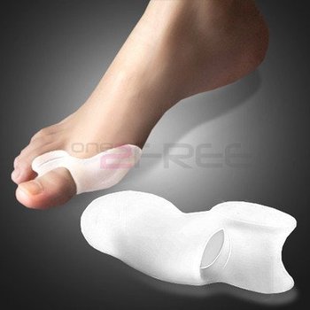 A bunion (Hallux Abducto Valgus) is sometimes described as a bump on the side of the big toe. However, the visible bump actually reflects changes in the bony framework in the front part of the foot. Instead of pointing straight ahead, the big toe leans towards the second toe, throwing the bones out of alignment and producing the ?bump? of the bunion. Bunions are a progressive disorder and gradually change the angle of the bones in your foot over the years. Symptoms usually occur in the later stages. The skin over the base of your big toe may become red and tender, and make wearing shoes painful. The bigger the bunion gets, the more it hurts to walk. Pressure from your big toe can force your second toe out of alignment, sometimes overlapping your third toe. Severe bunions can make it difficult to walk and you may develop arthritis.
Causes
No one single cause has been proven. There are a number of causes, and though shoes can exacerbate the problem, bunions do occur in societies that don?t wear them. We walk on the same type of ground all the time, whereas the human foot was actually designed to adapt to varying terrains. In a sense, a bunion is a type of repetitive strain injury. And like repetitive strain injury, some people are more prone to it than others. One theory, though it remains unproven, is that bunions are caused by one or both of the following. Because the foot wasn?t designed to constantly walk on a level surface, the ball of the big toe is slightly lower than the ball of the rest of your foot. When your foot meets the ground, the ball of the big toe is pushed up, and the big toe joint can?t bend as well as it was designed to. In order for the big toe joint to bend fully as you walk, your foot rolls slightly over to the side (this is also why people with hallux valgus often get hard skin). Also, if your midtarsal joint tends to move from side to side more than it does up and down, the arch in your foot collapses as your foot rolls in. This also makes you more prone to developing bunions. Such problems can be exacerbated by tight footwear. Slip-on shoes can make matters worse. Because they have to be tighter to stay on your feet, you automatically have less room for your toes. And with nothing to hold your foot in place, your toes often slide to the end where they?re exposed to lots of pressure. Likewise, high heels throw more weight onto the ball of the foot, putting your toes under further pressure. If you haven?t got a bunion by adulthood and you later develop one, there could be some underlying arthritis.
Symptoms
Bunions typically start out as a mild bump or outward bending of the big toe. Bunions at this stage are usually only a concern of appearance at this stage, and at this point they often don't hurt much. Over time, the ligaments that connect the bones of the toe stretch out, and the tendons attaching to the big toe gradually pull it farther and farther towards the second toe. Sometimes patients will find their first and second toes begin to press together too much, and they'll often get a painful corn between those toes. As the bunion progresses, the big toe may begin to ride on top of the second toe, or vice versa, creating a second deformity. Others will develop bump pain at the site of the bony enlargement on the side of the foot. A painful bursa may develop at that site. This is particularly true in tight shoes. Many patients also develop a painful callus beneath the foot. Capsulitis and other types of metatarsalgia may develop in the joints beneath these calluses, particularly in the second and third metatarsophalangeal joints (the joints in the ball of the foot). Over time, with the toe held in a crooked position for enough time, arthritis develops in the big toe joint. This will usually result in decreased range of motion of that joint (a condition known as "Hallux Limitis"), which as a result, often causes the patient to changes in the way a patient walks. Often the patient walks in an "out-toed", or duck-like, fashion, which very frequently causes secondary pain in the legs, knee, hip, and low back.
Diagnosis
Orthopaedic surgeons diagnose bunions on the basis of physical examination and weight bearing x-rays. Two angles are assessed, the intermetatarsal angle, that is between the first and second metatarsals (the bones that lead up to the base of the toes). If this angle exceeds 9? (the angle found in the healthy foot) it is abnormal and referred to as metatarsus primus varus. the hallux valgus angle, that is, the angle of the big toe as it drifts toward the small toe. An angle that exceeds 15? is considered to be a sign of pathology.
Non Surgical Treatment
Treatment options vary depending on the severity of your bunion and the amount of pain it causes you. Early treatment is best to decrease your risk of developing joint deformities. Conservative treatment Nonsurgical treatments that may relieve the pain and pressure of a bunion include changing shoes. Wear roomy, comfortable shoes that provide plenty of space for your toes. Padding and taping. Your Podiatrist can help you tape and pad your foot in a normal position. This can reduce stress on the bunion and alleviate your pain.(Obviously pending on footwear selection). Medications. Acetaminophen (Tylenol, others) can control the pain of a bunion. Your doctor may suggest nonsteroidal anti-inflammatory drugs (NSAIDs), such as ibuprofen (Advil, Motrin, others) or naproxen (Aleve), for relieving pain and reducing inflammation. Cortisone injections also can be helpful. But keep in mind that medications do not alleviate the actual cause of the pain. Physical therapy. The heating effect of ultrasound therapy or whirlpool baths can provide relief from the pain and inflammation of a bunion. Orthotics can help control abnormal movement of your foot, reducing your symptoms and preventing your bunion from getting worse. Over-the-counter arch supports can provide relief for some people, though others may require prescription orthotics. 
Surgical Treatment
There are a range of different surgeries that can be performed with the goal of realigning the joint and relieving pain ranging from shaving off part of the bone to cutting and realigning the bone with pins and screws. Depending on the surgery full recovery can take months and require you to stay off the foot. One new type of surgery, called a tightrope, involves attaching a wire to the bone to try and pull it back into alignment, but be wary of this procedure because there have not been any long-term outcome studies yet.
The Facts Regarding Over-Pronation
Overview
Over pronation is when there is excessive or too much movement of the foot. Over pronation is a very common cause of heel pain and general pain throughout the lower extremities. This condition can often be referred to as flat feet and causes you to walk on other parts of your foot, which is what leads to serious heel and foot pain.
Causes
In adults, the most common reason for the onset of Over-Pronation is a condition known as Post Tibial Tendonitis. This condition develops from repetitive stress on the main supporting tendon (Posterior Tibial Tendon) of the foot arch. As the body ages, ligaments and muscles can weaken. When this occurs the job of providing the majority of the support required by the foot arch is placed upon this tendon. Unfortunately, this tendon cannot bear the weight of this burden for too long. Eventually it fatigues under the added strain and in doing so the foot arch becomes progressively lower over a period of time.
Symptoms
Over-Pronation may cause pain in the heel of the foot, the foot arch, under the ball of the foot, in the ankle, knee, hip or back. The symptoms may be localized to one particular area of the foot or may be experienced in any number of combinations. Standing for long periods of time, walking and running may become difficult due to the additional stress and/or discomfort accompanied with these activities. Upon Visual Inspection, when standing the heels of the foot lean inward and one or both of the knee caps may turn inward.
Diagnosis
When you overpronate your foot rolls inwards causing the lower leg to rotate inwards too. It's thought that this increases stress on the soft tissues of the lower leg causing pain and inflammation, or more commonly known as shin splints.
Non Surgical Treatment
Fortunately, there are simple things you can do to cure and correct your overpronation issues. Certain exercises help. Pull your toes back using a rolled up towel. Roll your feet over a golf or tennis ball for a minute. And do calf raises by standing up and lifting up on your toes. These all help reposition the foot and strengthen the muscles and tendons necessary for proper support. Beyond that, simple adjustments to footwear will help immensely.
Surgical Treatment
Depending on the severity of your condition, your surgeon may recommend one or more treatment options. Ultimately, however, it's YOUR decision as to which makes the most sense to you. There are many resources available online and elsewhere for you to research the various options and make an informed decision.
Can I Take Care Of Severs Disease In The Home?
Overview
Sever's disease or calcaneal apophysitis, is the most common cause of heel pain in the growing athlete and is due to overuse and repetitive microtrauma of growth plates of the calcaneus in the heel. It occurs in children ages 7 to 15, with the majority of patients presenting between 10 and 12 years of age.
Causes
The cause of the pain in Severs disease is thought to be the tractional forces applied to the growth plate of the heel bone by the Achilles tendon at the rear of the heel bone and the plantar fascia just beneath the heel bone. This pulling force by the Achilles tendon on the growth plate is often aggravated by tight calf muscles and excessively pronated feet (i.e. feet that ?roll in? too far).
Symptoms
Unilateral or bilateral heel pain. Heel pain during physical exercise, especially activities that require running or jumping or are high impact. Pain is often worse after exercise. A tender swelling or bulge on the heel that is painful on touch. Limping. Calf muscle stiffness first thing in the morning.
Diagnosis
Your Podiatrist or Physiotherapist will assist in diagnosing the injury and the extent of the damage. From this, they will develop a management plan which may include rest or activity modification, soft tissue treatment such as massage and stretching, correction of biomechanics through heel raises or orthoses and the progression through a series of specific strengthening exercises.
Non Surgical Treatment
First, your child should cut down or stop any activity that causes heel pain. Apply ice to the injured heel for 20 minutes 3 times a day. If your child has a high arch, flat feet or bowed legs, your doctor may recommend orthotics, arch supports or heel cups. Your child should never go barefoot. If your child has severe heel pain, medicines such as acetaminophen (one brand name: Tylenol) or ibuprofen (some brand names. Advil, Motrin, Nuprin) may help.
What Are The Causes And Indications Of Achilles Tendon Rupture
Overview
 The Achilles tendon attaches the calf muscles in the leg to the heel bone. It is the largest yet most exposed tendon in the body. An Achilles tendon rupture injury is when the tendon fibres tear, causing symptoms of pain and loss of function. A rupture can be either partial or complete and treatment may involve surgery. Achilles tendon rupture is most common in weekend athletes trying to train too hard and is least common in well-trained professional athletes. The injury is more common in men than in women and the frequency of rupture increases over the age of 30 years.
Causes
Often an Achilles rupture can occur spontaneously without any prodromal symptoms. Unfortunately the first "pop" or "snap" that you experience is your Achilles tendon rupture. Achilles tendon rupture most commonly occurs in the middle-aged male athlete (the weekend warrior who is engaging in a pickup game of basketball, for example). Injury often occurs during recreational sports that require bursts of jumping, pivoting, and running. Most often these are tennis, racquetball, squash, basketball, soccer, softball and badminton. Achilles rupture can happen in the following situations. You make a forceful push-off with your foot while your knee is straightened by the powerful thigh muscles. One example might be starting a foot race or jumping. You suddenly trip or stumble, and your foot is thrust in front to break a fall, forcefully over stretching the tendon. You fall from a significant height. It does appear that previous history of Achilles tendonitis results in a degenerative tendon, which can grow weak and thin with age and lack of use. Then it becomes prone to injury or rupture. Certain illnesses (such as arthritis and diabetes) and medications (such as corticosteroids and some antibiotics) can also increase the risk of rupture.
Symptoms
It is important to know that pain at the back of the heel is not always due to Achilles tendon rupture. It may be due to bursitis (fluid accumulation in the heel due to repeated irritation) and tendonitis (pain along the Achilles tendon due to constant friction and irritation). The above disorders tend to improve with use of pain medications and rest, whereas Achilles tendon rupture requires surgery and/or a cast.
Diagnosis
A doctor will look at the type of physical activity you have been doing. He or she will then look at your foot, ankle and leg. An MRI may also be used. This is to help determine the severity of the tear and the extent of separation of the fibers.
Non Surgical Treatment
The other option is to allow your tendon to heal without surgery. In this case, you also need to wear a cast, splint, walking boot, or brace for 6-8 weeks. You also may have different exercises to do. If you are less active or have a chronic illness that prevents surgery, this option may be better for you. 
Surgical Treatment
Surgery will involve stitching the two ends of the tendon together, before placing the leg in a cast or brace. The advantage of having an operation is the reduced chance of the rupture reoccurring, however it will involve the risks associated with any surgical procedure, such as infection.
Prevention
Prevention centers on appropriate daily Achilles stretching and pre-activity warm-up. Maintain a continuous level of activity in your sport or work up gradually to full participation if you have been out of the sport for a period of time. Good overall muscle conditioning helps maintain a healthy tendon.
True Leg Length Discrepancy Tests
Overview
Surgery is another option. In some cases the longer extremity can be shortened, but a major shortening may weaken the muscles of the extremity. In growing children, lower extremities can also be equalized by a surgical procedure that stops the growth at one or two sites of the longer extremity, while leaving the remaining growth undisturbed. Your physician can tell you how much equalization can be attained by surgically halting one or more growth centers. The procedure is performed under X-ray control through very small incisions in the knee area. This procedure will not cause an immediate correction in length. Instead, the LLD will gradually decrease as the opposite extremity continues to grow and "catch up." Timing of the procedure is critical; the goal is to attain equal length of the extremities at skeletal maturity, usually in the mid- to late teens. Disadvantages of this option include the possibility of slight over-correction or under-correction of the LLD and the patient?s adult height will be less than if the shorter extremity had been lengthened. Correction of significant LLDs by this method may make a patient?s body look slightly disproportionate because of the shorter legs.
Causes
Leg discrepancy can develop from a medical issue in any portion of the femur or tibia. One leg may lengthen, but leg shortening is much more common. Factors that can cause leg length discrepancy include inherited growth deficiencies. Infections. A bone infection can cause delayed growth in the affected limb. Injury. If your child breaks a leg, it may be shorter once it heals. This is most likely to happen if the fracture or break was complicated, an open fracture, or an injury that affected the growth plate near the end of the bone. Alternatively, a break can cause bones to grow faster after healing, making a leg longer. Tumors. Legg-Calve-Perthes disease. This is a condition that affects the ball (femoral head) of the hip joint. The femoral head may be friable and damage easily, sometimes leading to shortening of the thigh bone. Hemihypertrophy. In children with this condition, one side of the body grows more quickly than the other. Vascular malformations. These are abnormal clusters of veins and arteries that can form close to the bone and stimulate growth. Juvenile arthritis. Inflammation from arthritis can stimulate growth in the affected leg and cause discrepancy.
Symptoms
Children whose limbs vary in length often experience difficulty using their arms or legs. They might have difficulty walking or using both arms to engage in everyday activities.
Diagnosis
Infants, children or adolescents suspected of having a limb-length condition should receive an evaluation at the first sign of difficulty in using their arms or legs. In many cases, signs are subtle and only noticeable in certain situations, such as when buying clothing or playing sports. Proper initial assessments by qualified pediatric orthopedic providers can reduce the likelihood of long-term complications and increase the likelihood that less invasive management will be effective. In most cases, very mild limb length discrepancies require no formal treatment at all.
Non Surgical Treatment
Treatment depends on the amount and cause of the leg length discrepancy as well as the age of your child. Typically, if the difference is less than 2 cm we don?t recommend immediate treatment. We may recommend that your child wear a heel lift in one shoe to make walking and running more comfortable. If the leg length discrepancy is more significant, your doctor may recommend surgery to shorten or lengthen a leg. The procedure used most often to shorten a leg is called epiphysiodesis.

Surgical Treatment
Leg shortening is employed when LLD is severe and when a patient has already reached skeletal maturity. The actual surgery is called an osteotomy , which entails the removal of a small section of bone in the tibia (shinbone) and sometimes the fibula as well, resulting in the loss of around an inch in total height. Leg lengthening is a difficult third option that has traditionally had a high complication rate. Recently, results have improved somewhat with the emergence of a technique known as callotasis , in which only the outer portion of the bone (the cortex ) is cut, (i.e. a corticotomy ). This allows the bone to be more easily lengthened by an external fixation device that is attached to either side of the cut bone with pins through the skin. The ?ex-fix,' as it is sometimes called, is gradually adjusted by an orthopaedic surgeon, and healing can occur at the same time that the leg is being distracted , or lengthened over time. Unlike epiphysiodesis, leg lengthening procedures can be performed at almost any skeletal or chronological age.
Surgery is another option. In some cases the longer extremity can be shortened, but a major shortening may weaken the muscles of the extremity. In growing children, lower extremities can also be equalized by a surgical procedure that stops the growth at one or two sites of the longer extremity, while leaving the remaining growth undisturbed. Your physician can tell you how much equalization can be attained by surgically halting one or more growth centers. The procedure is performed under X-ray control through very small incisions in the knee area. This procedure will not cause an immediate correction in length. Instead, the LLD will gradually decrease as the opposite extremity continues to grow and "catch up." Timing of the procedure is critical; the goal is to attain equal length of the extremities at skeletal maturity, usually in the mid- to late teens. Disadvantages of this option include the possibility of slight over-correction or under-correction of the LLD and the patient?s adult height will be less than if the shorter extremity had been lengthened. Correction of significant LLDs by this method may make a patient?s body look slightly disproportionate because of the shorter legs.
Causes
Leg discrepancy can develop from a medical issue in any portion of the femur or tibia. One leg may lengthen, but leg shortening is much more common. Factors that can cause leg length discrepancy include inherited growth deficiencies. Infections. A bone infection can cause delayed growth in the affected limb. Injury. If your child breaks a leg, it may be shorter once it heals. This is most likely to happen if the fracture or break was complicated, an open fracture, or an injury that affected the growth plate near the end of the bone. Alternatively, a break can cause bones to grow faster after healing, making a leg longer. Tumors. Legg-Calve-Perthes disease. This is a condition that affects the ball (femoral head) of the hip joint. The femoral head may be friable and damage easily, sometimes leading to shortening of the thigh bone. Hemihypertrophy. In children with this condition, one side of the body grows more quickly than the other. Vascular malformations. These are abnormal clusters of veins and arteries that can form close to the bone and stimulate growth. Juvenile arthritis. Inflammation from arthritis can stimulate growth in the affected leg and cause discrepancy.
Symptoms
Children whose limbs vary in length often experience difficulty using their arms or legs. They might have difficulty walking or using both arms to engage in everyday activities.
Diagnosis
Infants, children or adolescents suspected of having a limb-length condition should receive an evaluation at the first sign of difficulty in using their arms or legs. In many cases, signs are subtle and only noticeable in certain situations, such as when buying clothing or playing sports. Proper initial assessments by qualified pediatric orthopedic providers can reduce the likelihood of long-term complications and increase the likelihood that less invasive management will be effective. In most cases, very mild limb length discrepancies require no formal treatment at all.
Non Surgical Treatment
Treatment depends on the amount and cause of the leg length discrepancy as well as the age of your child. Typically, if the difference is less than 2 cm we don?t recommend immediate treatment. We may recommend that your child wear a heel lift in one shoe to make walking and running more comfortable. If the leg length discrepancy is more significant, your doctor may recommend surgery to shorten or lengthen a leg. The procedure used most often to shorten a leg is called epiphysiodesis.
Surgical Treatment
Leg shortening is employed when LLD is severe and when a patient has already reached skeletal maturity. The actual surgery is called an osteotomy , which entails the removal of a small section of bone in the tibia (shinbone) and sometimes the fibula as well, resulting in the loss of around an inch in total height. Leg lengthening is a difficult third option that has traditionally had a high complication rate. Recently, results have improved somewhat with the emergence of a technique known as callotasis , in which only the outer portion of the bone (the cortex ) is cut, (i.e. a corticotomy ). This allows the bone to be more easily lengthened by an external fixation device that is attached to either side of the cut bone with pins through the skin. The ?ex-fix,' as it is sometimes called, is gradually adjusted by an orthopaedic surgeon, and healing can occur at the same time that the leg is being distracted , or lengthened over time. Unlike epiphysiodesis, leg lengthening procedures can be performed at almost any skeletal or chronological age.